Long-term live cell imaging and cardiomyocyte function evaluation using ImageXpress Micro
Live cell imaging technology is a technology that has been developed in recent years to observe cell morphological changes and protein expression in a state where cells are close to physiological. This technique can avoid the traditional method of using fixed cells or tissues to fix cells. The resulting changes in cell morphology and structural changes can more accurately reflect the characteristics of cells and are therefore widely recognized. The MD ImageXpress Micro High Connotation System has a wide range of features for live cell observations for up to several days, and is highly flexible and scalable thanks to MetaXpress's high-content analysis software based on the well-known MD software product MetaMorph. In addition to live cell imaging, it is possible to evaluate the physiological and pharmacological properties of certain cells, such as cardiomyocytes.
Long-term imaging of nerve cells
Nerve cells are recognized as sensitive and fragile cells and are sensitive to the external environment. Long-term cultivation and observation of living nerve cells is difficult. Long-term observation and culture of nerve cells can be achieved using the ImageXpress Micro system.
method:
Tissue blocks of approximately 1 cubic millimeter in size were obtained from the hippocampus of neonatal rats. Transfer to a centrifuge tube containing 0.25% trypsin for digestion for 30 to 30 minutes at 37 ° C in a 5% CO 2 incubator. The digestion was terminated by adding a culture solution containing fetal bovine serum, and the cells were resuspended by centrifugation and plated in a 96-well plate at 0.6 × 10 5 /ml. After 24 h, it was changed to Neurolbasal Medium/B27 serum-free medium, and then changed once every 2~3 d. Primary nerve cells were obtained after 7 days.
Image acquisition:
The sample was placed in the MD ImageXpress Micro high-content system with environmental control function, and the sample was focused by laser autofocus. The phase difference image was taken every 5 minutes under the 10X objective lens. The results are as follows:


summary:
ImageXpress Micro's environmental control system maintains the temperature, humidity and CO 2 conditions required by the cells. The temperature provides temperature protection for the enzyme activity of the cells, ensuring the physiology of the cells, and maintaining the humidity to ensure that the extracellular fluid does not evaporate. Cell dehydration caused by an increase in extracellular osmotic pressure is avoided, and CO 2 can maintain the pH of the extracellular fluid.
In addition to maintaining the living environment of cells, during the long-term observation of sensitive and vulnerable cells (such as nerve cells), it is most important to reduce the stimulation of the cells and maintain the activity of the cells as much as possible. ImageXpress Micro uses laser autofocus technology. During the focusing process, the cells do not need to be exposed to light, thus avoiding the photochemical reaction and temperature rise in the cells, which can maintain the active state of the cells. Therefore, the above two points are indispensable during the observation of living cells for a long time.
Functional evaluation of cardiac stem cell
The software system used by ImageXpress Micro is powerful and has unlimited extensions. Therefore, in addition to the long-term observation of living cells and the analysis of cell morphology and protein expression, ImageXpress Micro can also evaluate cell cytology.
Stem cell research is currently a hot topic in biological research. The use of human stem cells (such as cardiac stem cells) can speed up the research, while also reflecting the function of human cells, and speeding up the entry of drugs into the clinic. The iPS-derived cardiomyocytes are particularly attractive for their expression of ion channels, spontaneous rhythm characteristics, and characteristics similar to those of primary cardiomyocytes. This cell allows for the study of myocardial cell characteristics and shortens the time and clinical screening of drugs. The period of the previous experiment.
method:
Cellular cells derived from Cellular Dynamics iPS were plated in 96-well plates and incubated in culture for 2-7 days, incubated with Calcein AM for 10 minutes, discarded after culture, and added with different concentrations of agonist. And inhibitors.
Image acquisition and results:
The maturation of cardiomyocytes is determined by the spontaneous rhythmic contraction of the cells. The rhythmic contraction of cells can be detected by electrophysiological methods. But the electrophysiological method is relatively complicated, so we replaced it with automatic analysis of fluorescent images.
First, we can obtain a set of images of cell rhythmic contraction by time-lapse or real-time shooting.

MetaXpress automatically analyzes myocardial cell beating
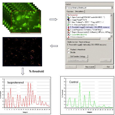
FIG. Beating cardiomyocytes obtained a set of images generated stack of images, obtained using the extended function module automatic analyzer Journal adjacent time difference between the two points of the image, determines a beat frequency and amplitude of the myocardial cells using the function The counter in the module automatically calculates the frequency and amplitude of myocardial beats. The above functions are automatically completed in the function module of the Journal extension, and directly report the myocardial cell beat frequency and amplitude information.
Myocardial beat pattern detection
We used the classical positive drugs isoproterenol, adrenaline, and caffeine as stimulators to detect the myocardial beat pattern and the function of the myocardial beat analysis module using the known cardiomyocyte inhibitor acetylcholine.
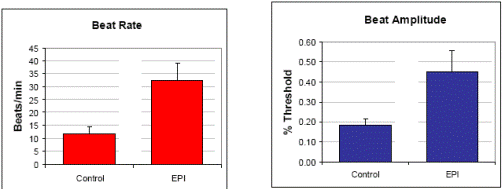
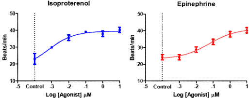

Changes in the frequency of myocardial cell beats under different concentrations of four drugs. The spontaneous rhythm of the cells varies from 20 to 40 beats per minute. The myocardial beat frequency was stable within 5 to 60 minutes after administration, so the above results were all 10 minutes after administration.
summary:
ImageXpress Micro's highly flexible system enables automated imaging of cardiomyocytes, and its MetaXpress software is extremely flexible, with its Journal function allowing for arbitrary expansion based on image and experimental requirements, in addition to cardiomyocyte physiology evaluation. In addition, more than 1000 functions can be expanded.
Polypropyl carbonate is soluble in polar solvents such as ketones, ethyl acetate, dichloromethanes and chlorohydrocarbons, and insoluble in alcohols, water and aliphatic hydrocarbons. It also forms a stable emulsion in the water. PPC allows gases like oxygen to diffuse through it. The glass temperature (Tg) is between 25 and 45℃, and PPC adhesives are amorphous. The glass temperature of PPC is slightly higher than that of polyethyl carbonate (PEC).
Poly Propylene Carbonate Ppc,100% Recycled Ppc,Bio-Based Ceramic Binder Ppc,Electroceramics Application Ppc
Xingbang High Molecular Materials Co., Ltd. , https://www.chemicaladditive.com