xCELLigence system for real-time monitoring of low toxicity X-tremeGENE DNA transfection experiments
Foreword
X-tremeGENE DNA Transfection Reagent is a non-liposome, multi-component transfection reagent that has been shown to efficiently transfect a wide variety of cells. X-tremeGENE 9 and X-tremeGENE HP can be transfected in serum-free conditions with low cytotoxicity and no need to change media after transfection.
It is very important to reduce the off-target effect of the transfection reagent itself:
â– Guarantee high expression rate of transfected target genes for a long time
â– Reduce the number of dead cells and the proteases they release, and high expression of recombinant proteins that are not digested.
â– Obtain results related to transgenic target organisms
In this study, the cytotoxicity and transfection efficiency of X-tremeGENE 9 and X-tremeGENE HPTransfection Reagent were tested on Hela cells and compared with other reagents.
step
â– Cytotoxicity The xCELLigence RTCA MP instrument is used to detect cell electrical activity, including cell adhesion, stretching, proliferation, death, and other morphological changes, by measuring electrical impedance for a long time, without labeling. The newly-launched consumable E-plate VIEW 96 has a 0.5mm transparent window in the lower part of each hole for visual inspection.
â– Roche Automated Image Capture and Analysis System Cellavista, a fully automated, high-throughput, precision cell image analysis workstation that combines the advantages of visible light and fluorescence imaging to observe cell morphological changes and detect transfection efficiencies in experiments.
â– Cellular metabolic activity can be assayed by WST-1 cell proliferation kit to quantify the metabolic activity of active cells.
result
Cytotoxicity
■Continuous monitoring of the xCELLigence RTCA MP instrument showed that the GFP expression plasmid was transfected on Hela cells using X-tremeGENE 9 and X-tremeGENE HP Transfection Reagent, similar to the Cell Index (CI) values ​​of the untreated control group. ( Figure 1 ). The other reagents showed a significant decrease in cell index values ​​after 5 hours of transfection, indicating the presence of cytotoxicity.

Figure 1: Cell index change plots detected by xCELLigence after transfection of GFP expression plasmids on HeLa cells using X-tremeGENE 9 and X-tremeGENE HP Transfection Reagent and their transfection reagents.
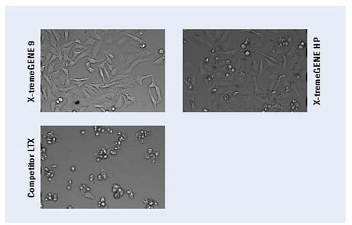
Figure 2: Morphology of Hela cells after transfection. Visible light image obtained by Cellavis instrument (10x magnification)
â– Cellavist visualized the morphology of the cells and obtained a visible light image after 24 hours of transfection. X-tremeGENE 9 and X-tremeGENE HP-transfected Hela cells are in a healthy state, while other reagent groups have a large number of dead cells (Figure 2).
â– End point analysis, cell activity was determined by cell proliferation kit WST-1 analysis after approximately 72 h of transfection (Fig. 3). Compared with X-tremeGENE 9 and X-tremeGENE HP Transfection Reagent, other reagents significantly reduced metabolic activity after transfecting cells, showing cytotoxic effects.
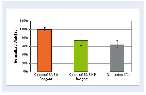
Figure 3: Comparison of cell viability after conversion of Roche X-tremeGENE 9 and X-tremeGENE HP with its reagents. Values ​​were determined by the WST-1 kit and normalized to the X-tremeGENE 9 results.
Transfection efficiency
â– For the number of cells expressing GFP after transfection with X-tremeGENE 9 and X-tremeGENE HP Transfection Reagent and other reagents, Cellavista recorded fluorescent images at the indicated time points (Fig. 4). The Cellavista image analysis software was used to quantify the GFP-positive cell area (fluorescence image) and the total cell area (corresponding visible light image, not shown). See Figure 5 for data. During the observation process, X-tremeGENE reagents have higher transfection efficiency.

Figure 4: X-tremeGENE Transfection Reagent transfected cells express large amounts of GFP. After 48 h of transfection, cells recorded by Cellavista expressed fluorescent images of GFP (10-fold magnification).
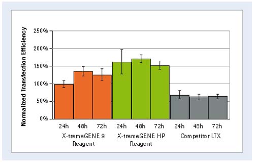
Figure 5: Quantification of GFP expression levels. The Cellavista analysis software counted the percentage of transfection efficiency after 24 h, 48 h, and 72 h after transfection (normalized by the value obtained after X-tremeGENE 9 Transfection Reagent transfection for 24 h).
to sum up
X-tremeGENE 9 and X-tremeGENE HP Transfection Reagent demonstrated low cytotoxicity and high DNA transfection efficiency in Hela cell line assays. The xCELLigence RTCA MP is also ideal for instruments that detect reagent-specific effects in real time.
Materials and Method
Cell culture
Hela cells (ATCC) were seeded in a suitable cell culture medium containing additional components and cultured at 37 ° C in a 5% CO 2 humidified environment. Cells were trypsinized, washed and seeded on E-Plate VIEW 96.
Transfection
24 hours after the inoculation, the GFP-pcDNA3.1 plasmid of the CMV promoter was transfected with three transfection reagents X-tremeGENE 9, X-tremeGENE HP and other reagents (LTX and L2K), respectively. Transfection According to the instructions in the instruction manual, the ratio of optimal DNA to transfection reagent recommended in each instruction is used. All experiments were repeated three times.
Real-time cell analysis
The xCELLigence RTCA MP instrument continuously monitors the overall cell status of the experiment (from cell inoculation to three days after transfection). The value of 50 μl of cell culture medium per well was determined as the background electrical impedance. The final volume per well was adjusted to 100 μl and the density was 4000 Hela cells. After inoculation, the electrical impedance was measured at fixed time intervals. Cell index (CI) values ​​were normalized at the time of transfection.
Automatic imaging
The Cellavista system high-throughput microscope can be operated at specified time points during the experiment. The xCELLigence system measurement can be briefly paused and the E-Plate VIEW 96 can be converted into the Cellavista system. Visible and fluorescent images of all 96-well plates were obtained with a 10x objective. Transfection efficiency can be obtained using Cellavista software analysis. Once the image is acquired, the E-Plate VIEW 96 returns to the xCELLigence RTCA MP instrument and the cell resistance measurement can be restarted.
WST-1 analysis
Cell viability can be determined at the end of the experiment using the WST-1 Cell Proliferation Kit (Roche Applied Science) (72 hours after transfection), see instructions.
English original text:
Real-time monitoring of low cytotoxic X-tremeGENE DNA Transfection Reagents using the xCELLigence System
Vaseline Gauze,Vaseline Gauze Pads,Vaseline Coated Gauze,Sterile Vaseline Gauze
Henan Anbang Medical Supplies Co., Ltd. , https://www.anbangmedical.com